


In vitro and in vivo antifungal activity of extracts from Jamaica cherry Muntingia calabura l. leaves
- Faculty of Chemical Engineering and Food Technology, Nong Lam University, Vietnam
- Faculty of Bioscience Engineering, Ghent University, Belgium
Abstract
Previous studies on M. calabura (Jamaica cherry) leaves reported the presence of polyphenols, flavonoids, glycosides, and tannins. However, the deep research focus on the antifungal activity of Jamaica cherry leaf extract (JCLE) is still limited. Hence, the current study was conducted on JCLE samples by using various solvents, i.e., 70% ethanol, 70% methanol, and 70% acetone, and then evaluating their antifungal activity. Analytical results showed that JCLE could be used as a natural antifungal potential agent against mold-causing spoilage in fruits. Specifically, the ethanolic extract had a strong inhibition of Colletotrichum gloeosporioides and Lasiodiplodia theobromae. While the methanolic and acetonic extracts prevented sharply the growth of Alternaria alternata and Fusarium equiseti, respectively. Generally, the ethanolic extract was able to inhibit the various mold strains better than the other solvents. Furthermore, the results showed that the percentage inhibition of radial growth (PIRG) of JCLE increased gradually as the concentration increased from 4000 to 40000 mg GAE/kg. The highest data obtained for C. gloeosporioides, A. alternata, F. equiseti, and L. theobromae were 53.29%, 57.03%, 54.81%, and 75.03%, respectively. In vivo experiment, the samples were treated with and without JCLE (the control sample) occuring the black spots caused by C. gloeosporioides on mango’s surface after 21 days and 30 days of storage at 12 ± 2 °C, respectively. Whereas the TSS and TA values of both samples were similar. While the visual appearance of the control was more yellow compared to the samples treated with JCLE. In conclusion, JCLE was able to significantly delay the development of pathogenic mold, though it quite inhibited the maturation of mango fruit. Further studies may be needed to evaluate the phtychemical profile of M. calabura extract to gain a deeper understanding of its antifugal activity.
INTRODUCTION
One of the most critical factors that deteriorated the quality and quantity of fresh fruits during postharvest is the growth of the fungal pathogen. The famous fungi, such as Alternaria spp, Lasiodiplodia spp, Colletotrichum spp and Fusarium spp, which caused rots and anthracnose1. The fungicides are used as a solution to inhibit fungal growth and to reduce fruit spoilage, but their adverse effects on the environment and human health also could be concern issues. So far, the scientists have focused on the research about the bioactive compounds (e.g., polyphenol, alkaloids, sterols, essential oils…) available in the extracts from plants and plant-based by-products with their antimicrobial ability2, 3, 4. These results show the potential to apply Jamica cherry extracts post-harvest, and these extracts possibly replace the chemicals due to the safety in usage and the friendliness to the environment.
Muntingia calabura L. (M. calabura), a shrub originally from the American continent, is well-known as the “Jamaica cherry” or another name as “Trứng cá.” It is widely cultivated in tropical regions, especially in several Asian countries, including Vietnam. M. calabura, especially leaves, stems, and roots, have been documented to have traditional medicinal usage in various modes of applications5. Previous studies on M. calabura leaves reported the presence of polyphenol, flavonoid, glycoside, and tannin6, 7. However, the research specifically focusing on the antifungal activity of M. calabura leaves extract is still limited. Therefore, this study evaluated the effect of M. calabura L, leave extract on the growth of several spoilage fungi such as Alternaria alternata, Lasiodiplodia theobromae, Colletotrichum gloeosporioides, and Fusarium equiseti. The leaves were extracted with different solvents, including ethanol, methanol, and acetone, to collect the leave extracts that were applied to the in vitro and in vivo antifungal activity based on the assessment of the fungal growth zone.
On the other hand, Mango is one of the most important tropical fruits in Vietnam. However, it is highly susceptible to various pathogens of which anthracnose caused by C. gloeosporioides is the most common2. Therefore, the aim of this study is preliminary evaluate the effect of M. calabura L, leave extract on the growth of this strain.
MATERIALS AND METHODS
Materials
Chemicals and culture media
Ethanol (EtOH) 99.5%, methanol (MeOH) 99.5%, acetone (Ace) 99.5%, sodium chloride (NaCl), and sodium carbonate (NaCO) were obtained from Xilong company (China). The dimethyl sulfoxide (DMSO), gallic acid, and Folin - Ciocalteu reagent were purchased from Sigma-Aldrich (USA). The potato Dextrose Agar (PDA) was purchased from Himedia company (India).
Preparation of the extract
The Jamaica cherry leaves were collected in Thu Duc City, Ho Chi Minh City, Vietnam. The fresh leaves (4 – 15 cm in length and 1 – 6 cm in width) without pest and deterioration were selected, washed with tap water, and dried at 55 ± 2 in a hot air oven till moisture content ≤ 10%. Then, the dried leaves were ground and sieved through a 0.5 mm sieve to collect the powdery homogeneous sample. The powdery sample was stored in an aluminum foil Ziplock bag at room temperature (30 ± 2 C).
The extraction was carried out by soaking the dried plant powder (30 g) in 300 mL of the various solvents (i.e., ethanol 70%, methanol 70%, and acetone 70%) for 48 hours at room temperature (30 ± 2 C), followed by centrifugation (Hermle Z206 A, Germany) at 5000 rpm for 5 mins and filtration through Whatman no1 filter paper. The residue was then re-extracted with 150 ml of solvent with the same steps. The combined supernatant was concentrated in a rotary vacuum evaporator (RE-100D, Phoenix Instrument, Germany) at 50 until the solvent completely evaporated to obtain the dried extracts. The evaporation period was terminated when the collecting flask has not been dropped with any solvent for 5 minutes which indicated the solvent minimally present in a sample. The extract after evaporating was called JCLEC. The sample were stored in a fridge at 4-6 .
Fresh mango fruits
Mango (Mangifera indica L) is well-known as the “Cat Hoa Loc” mango. It was purchased in Dinh Quan District, Dong Nai Province, Vietnam. The fruit was harvested at the commercial maturity stage (based on skin color and firmness). The selected fruits were fresh, without defects of physical injuries and fungal injections. Each fruit weighs approximately 400 – 500 g with a soluble solids content of 6 – 7°Brix, total acid content of 0.90 – 1.00% and colorimetric values of L* from 63.04 to 65.23, a* from -15.15 to -12.11, and b* from 30.33 to 32.57. Fruits were washed with tap water and surface soaked with 100 mg/kg chlorine for 3 - 5 mins and dried at room temperature (30 ± 2 C) for further experiments.
Pathogenic fungus
The fungal strains as Colletotrichum gloeosporioides, Alternaria alternata, Lasiodiplodia theobromae, and Fusarium equiseti were isolated from mango and supplied by the Southern Horticultural Research Institute, Vietnam. These strains were stored at 4 in an agar tube containing mycelium slant and covered with a layer of sterile paraffin liquid to prevent dehydration of the agar. These strains were cultured on potato dextrose agar (PDA) at room temperature (30 ± 2 C) for 14 days (A. alternata), 7 days (C. gloeosporioides and F. equiseti), 3 days (L. theobromae) to create the filamentous form (growth zone) for the experiments. The culture method was followed by Estrada et al. (2000) with slight modifications8.
Experimental design
Effects of extraction solvents on TPC, recovery yield and antifungal activity of the obtained extracts from Jamaica cherry leaves
The JCLEC was carried out according to the procedure described in section 2.1.2, wherein the solvents such as ethanol 70%, methanol 70%, and acetone 70% (v/v) which were used to extract JCLE. The JCLECs were determined TPC using Folin – Ciocalteu assay and antifungal activity using the percentage inhibition of radial growth. This experiment was carried out four times.
The extracts were dissolved in dimethyl sulfoxide (DMSO) 5% (v/v) to reach a final concentration of 100 mg dry matter/g and then filtered with 0.45 μm membrane filters for sterilization. Then, the solution was well-mixed with the sterilized potato dextrose agar (PDA) media at a temperature of 55 ± 2 . The mixed solution was transferred to a petri dish (90 cm), solidified, and stored in a fridge. Similarly, the control sample was prepared by substituting the extract with DMSO of 5%. Then, the filamentous fungus (supplied in section 2.1.4) with a 6 mm diameter piece was placed in the center of these dishes. Next, these samples were incubated at room temperature (30 ± 2 C) at different times depending on the specific strain such as 192 hours for C. gloeosporioides, 240 hours for A. alternata, 288 hours for F. equiseti, and 48 hours for L. theobromae. The diameter of the filamentous form (growth zone) was measured by the digital caliper (Mitutoyo, Japan).
In vitro assessment antifungal activity of ethanolic from Jamaica cherry leaves under various the concentration
The JCLEC was prepared similarly to the processing in section 2.1.2. Next, JCLEC was dissolved in dimethyl sulfoxide (DMSO) of 5% (v/v) and added to the sterilized PDA media to obtain a final concentration of 4000; 10000; 20000 and 40000 (mg GAE/kg) The sample continues to be completed according to section 2.2.1. This experiment was carried out four times.
In vivo evaluation of antifungal activity of Jamaica cherry leaves extract (JCLE) on mango storage.
Firstly, mango fruits were cleaned with tap water. Next, the mango’s surface was wiped with 70% ethanol. Then, each fruit was inflicted with a 1 mm deep wound by 0.3 mm outer diameter of a sterile hypodermic needle. Putting a 6 mm - diameter piece of C. gloeosporioides on the wound. Dissolving the JCLE in distilled water (v/v) to obtain a final concentration of 40000 mg GAE/kg. The mango fruits were placed on the sterilized tray. Spraying with the JCLE solution until the surface of the fruits was wet and leaked on the tray. The sterile distilled water was used as a negative control. The fruits were incubated at room temperature (30 ± 2 C) for 2 hours and stored in a perforated carton box (size 45 cm x 35 cm x 15 cm) at 12 ± 2 , RH of 85 – 90%. Each box contained ten fruits. The experiment was repeated twice. The first day of the storage period was day 0. The growth of C. gloeosporioides was recorded after each three days in 33 days.
Analytical methods
Determination of total phenolic content (TPC)
The method was described by Phuong et al, (2020)9. Briefly, 1 mL of bi-distilled water, 1 mL extract and 0.5 mL Folin – Ciocalteu reagent 10% (v/v) was mixed well in a glass test tube for 6 mins. Then, 1.5 mL sodium carbonate solution 20% (v/v) and 1 mL of bi-distilled water was added into and mixed well. The samples were then incubated for 120 min at room temperature (30 ± 2C) in dark before measuring the absorbance at 760 nm (GENESYS 10S UV-VIS, Thermo Scientific, USA). The TPC value was expressed as gallic acid equivalents mg GAE / kg dry matter.
Determination of inhibition percentage based on growth diameter
The percentage inhibition of radial growth (PIRG) was determined at the end of the incubation time according to Al-Hetar et al., (2011)10.
Where: % PIRG is the percentage inhibition of radial growth of the experimental treatment.
% PIRGo is the percentage inhibition of radial growth of the DMSO treatment.
D is the diameter of the filamentous form (growth zone) on the control plate (e.g., distilled water).
D is the diameter of the filamentous form (growth zone) on the plates containing the extracts.
D is the diameter of the filamentous form (growth zone) on the plates containing DMSO.
Color measurement
The color of the mango fruits was measured by using a Chromameter CR – 400 (Konica Minolta, Japan).
Titratable Acidity (TA)
The method was carried out according to ISO 750:199811. An aquilot,10 mL of mango juice was diluted with distilled water up to 250 mL and filtered by filter paper (New Start, China). 25 mL of the solution was titrated with 0.1 N NaOH and 1% phenolphthalein solution as indicator. The experiment was replicated 3 times. Total acidity was calculated by the following equation:
Where: V: Volume of titrated NaOH 0.1 N (mL), V: Volume of titrated aliquot, V: Volume of the sample, c: molarity of NaOH solution (0.1 N), K: corresponding citric acid coefficient (0.064)
Total soluble solids content (TSS)
The total soluble solid content was measured by hand refractometer (ATAGO 0.0~33.0% Brix, Japan) as °Brix in 0.1% graduations.
Statistical analysis
Stat-graphics software and Microsoft Excel 2010 software were used to process the data. The data obtained were presented as means ± standard deviation. The results were analyzed statistically using one-way analysis of variance method (ANOVA) and the method currently being used to discriminate among the means is Fisher's least significant difference (LSD) procedure (p ≤ 0.05).
RESULT AND DISCUSSION
Effects of extraction solvents on antifungal activity of the obtained extracts from Jamaica cherry leaves
The antifugal activity versus solvents
The DMSO did not affect the growth of the mold, and it indicated that the antifungal activity result of JCLE was not interfered with by DMSO. In contrast, Table 2 showed the JCLE inhibited the growth of fungi. Generally, the solvents such as ethanol and method gave JCLE's antifungal activities higher than acetone. In most samples, this material was against C. gloeosporioides, F. equiseti, and A. alternata lower than L. theobromae.
The percentage inhibition of radial growth of three solvents for C. gloeosporioides were 40.02%, 38.95%, and 35.81%, respectively; L. theobromae were 62.06%, 59.85%, and 58.47 %, respectively. The antifungal activity of JCLE on C. gloeosporioides and L. theobromae decreased in the following order: 70% ethanol > 70% methanol > 70% acetone. Regarding the A. alternata, the antifungal activity of JCLE gradually reduced from 70% methanol (48.71%) to 70% ethanol (46.24%), and 70% acetone (37.15%) after 240 hours of incubation. The inhibitory effect on mycelium growth of 3 solvents was statistically significant at the confidence level α < 0.05. Extraction by using methanol gave the best results in inhibiting A. alternata. For the F. equiseti, PIRG reached the highest at 70% acetone (45.17%) after 288 hours of incubation. Its antifungal ability deteriorated in the following order 70% methanol and 70% ethanol, with the specific values were 40.91%, and 37.95% at the same time, respectively. However, these values were not statistically significant at the confidence level α < 0.05.
This experiment showed that each strain was affected differently under the various solvents, which agreed with the other authors12, 13, 14, 15. Another different study revealed that flavonoids is a subclass of polyphenol may inhibit fungi growth by rupturing plasma membranes and decreasing cell wall production and cell division16. Moreover, the plant extract or phenolic compound has demonstrated an impact on bacteria growth17, 18 by reacting with protein, inhibiting bacterial cell wall formation, and changing the membrane permeability. The interaction between plant extract and mold may be according to a similar mechanism.
The previous studies had shown that the variation in solvent polarity gave a significant difference in TPC in the plant extract. Therefore, the change in TPC of JCLE in this current study due to the various solvents could not be ruled out19, 20. The TPC of JCLE increased in the following order: methanol 70%, ethanol 70%, and acetone 70% (Table 1), resulting in a PIRG of an ethanolic extract that was higher than a methanolic extract. However, as the argument before stated, the highest PIRG value was different based on the mold strains and solvents. As a result, the antifungal activity of JCLE may come from the various compounds, not only polyphenols. Previous studies found alkaloids and steroids in M. calabura leaves7, 21, 22. The other studies displayed that these compounds limited the growth of mold. For instance, alkaloids affect antifungal activity by obstructing enzyme function23. While steroids have lipophilic features that can prevent fungus from germinating spores24.
The antifugal activity versus incubation time
Moreover, the data in Table 1 also enumerated the antifugal activity of JCLE just had a high impact at a certain incubation time. For instance, PIRG of C. gloeosporioides rose from 50.02% to 65.27% when the incubation time from 48 to 96 hours. However, the incubation time expanded to 192 hours, the antifugal activity of JCLE dropped to 38.95%. The other strains, PIRG was highest at 96 hours for C. gloeosporioides, at 192 hours for A. alternata and F. equiseti. The reason could be the bioactive compounds in JCLE need a certain time to diffuse from agar media to fungus to inhibiting them, resulted in the increase in PIRG. However, because of the incubation long time, the fungus strains gradually adapting to the JCLE, hence, the antifungal activity of JCLE was reduced.
From the above initial survey results, ethanol extraction was selected for the following experiments because of its safety and economic relevance.
The TPC of JCLE under various solvents
|
Solvent |
TPC (mg GAE/kg) |
|
Acetone 70% |
65760A ± 2.45 |
|
Methanol 70% |
47110B ± 1.46 |
|
Ethanol 70% |
51920B ± 4.94 |
Effect of extraction solvents on PIRG of JCLE under various solvents
|
Time (hours) |
Solvents | |||
|
5% DMSO |
70% Acetone |
70% Methanol |
70% Ethanol | |
|
| ||||
|
48 |
00.00D ± 0.00 |
32.51C ± 1.23 |
50.02A ± 0.58 |
45.33B ± 1.20 |
|
96 |
00.00D ± 0.00 |
60.53B ± 0.90 |
65.27A ± 0.95 |
54.31C ± 0.18 |
|
144 |
00.00C ± 0.00 |
47.71B ± 0.61 |
48.44B ± 0.84 |
50.05A ± 1.20 |
|
192 |
00.00C ± 0.00 |
35.81B ± 2.69 |
38.95A ± 1.32 |
40.02A ± 1.16 |
|
| ||||
|
48 |
00.00C ± 0.00 |
23.00B ± 0.76 |
38.90A ± 1.21 |
37.91A ± 1.69 |
|
96 |
00.00C ± 0.00 |
39.12B ± 2.19 |
47.14A ± 0.82 |
46.93A ± 0.62 |
|
144 |
00.00C ± 0.00 |
40.05B ± 2.55 |
47.81A ± 1.13 |
48.78A ± 1.01 |
|
192 |
00.00C ± 0.00 |
40.57B ± 1.56 |
48.31A ± 2.62 |
48.10A ± 0.30 |
|
240 |
00.00D ± 0.00 |
37.15C ± 1.32 |
48.71A ± 1.77 |
46.24B ± 1.02 |
|
| ||||
|
48 |
00.00C ± 0.00 |
42.61A ± 2.98 |
37.63B ± 2.37 |
35.83B ± 2.81 |
|
96 |
00.00C ± 0.00 |
51.09A ± 0.44 |
41.78B ± 2.38 |
41.89B ± 0.77 |
|
144 |
00.00C ± 0.00 |
53.03A ± 0.95 |
44.73B ± 0.77 |
45.22B ± 1.00 |
|
192 |
00.00D ± 0.00 |
57.14A ± 1.50 |
50.89B ± 0.93 |
44.88C ± 1.11 |
|
240 |
00.00D ± 0.00 |
57.87A ± 1.16 |
46.85B ± 0.53 |
45.44C ± 0.97 |
|
288 |
00.00D ± 0.00 |
45.17A ± 0.61 |
40.91B ± 0.67 |
37.95C ± 0.36 |
|
| ||||
|
24 |
00.00C ± 0.00 |
53.35B ± 2.80 |
52.57B ± 4.42 |
59.67A ± 4.25 |
|
48 |
00.00D ± 0.00 |
58.47C ± 0.70 |
59.85B ± 0.55 |
62.06A ± 0.96 |
In vitro effect of concentration of ethanolic extract on antifungal activity
In the tested TPC range, the percentage inhibition of radial was statistically significant with confidence α < 0.05 in different concentrations (Table 3). For instance, mycelial growth decreased when the TPC value increased from 4000 to 40000 mg GAE/kg. Under this data range, the PIRG value achieved from 38.15 to 53.29%, respectively (C. gloeosporioides); 45.69% to 57.03%, respectively (A. alternata); 37.98% to 54.81%, respectively (F. equiseti); and 61.48% to 75.03%, respectively (L. theobromae). Table 2 also showed the fungal growth decreased in the following order L. theobromae > A. alternata> F. equiseti > C. gloeosporioides. For instance, the PIRG value of these strains after 48 hours of incubation was 75.03%, 9.95%, 30.35%, and 15.69%, respectively.
The plant extracts had a high content of phenolic compounds that display great antifungal activity. At a concentration of 3000 mg GAE/kg, Gómez-Maldonado et al. (2020) found an extraction from the Manila kernel that could completely inhibit C. brevisporum. The other studies also showed the Silene armeria L extract limited the growth of F. solani (61.3%) and C. capsici (53.0%) at 1500 mg GAE/kg25. Another study reported that the F. oxysporum could be prevented by 42.2% using ethanolic extracts of Achillea santolina of 1000 mg GAE/kg26. Therefore, the JCELC dosage of 40000 mg GAE/kg was chosen as this study's result.
Effect of concentration of Jamaica cherry leaves extract on antifungal activity (
|
Time (hours) |
The dosage (mg GAE/kg) | |||
|
4000 |
10000 |
20000 |
40000 | |
|
| ||||
|
48 |
2.54D ± 1.06 |
4.61C ± 0.34 |
7.40B ± 0.89 |
15.69A ± 0.88 |
|
96 |
29.62C ± 2.69 |
31.67C ± 3.12 |
35.29B ± 1.29 |
41.97A ± 2.31 |
|
144 |
35.10C ± 0.18 |
39.55B ± 0.99 |
40.83B ± 2.55 |
48.07A ± 1.47 |
|
192 |
38.15C ± 1.86 |
44.62B ± 0.75 |
44.98B ± 0.42 |
53.29A ± 1.20 |
|
| ||||
|
48 |
2.44C ± 0.53 |
6.80B ± 1.08 |
9.19A ± 1.33 |
9.95A ± 0.80 |
|
96 |
25.71B ± 2.26 |
25.30B ± 3.02 |
27.02B ± 3.69 |
31.46A ± 2.46 |
|
144 |
21.91C ± 1.57 |
25.93B ± 1.81 |
26.12B ± 1.39 |
35.56A ± 0.97 |
|
192 |
17.08C ± 2.77 |
26.62B ± 2.94 |
28.80B ± 1.97 |
36.42A ± 2.53 |
|
240 |
45.69C ± 0.42 |
46.92C ± 2.47 |
53.49B ± 0.85 |
57.03A ± 0.40 |
|
| ||||
|
48 |
21.53C ± 0.88 |
23.07BC ± 2.59 |
24.51B ± 2.51 |
30.35A ± 1.75 |
|
96 |
25.71B ± 2.26 |
25.30B ± 3.02 |
27.02B ± 3.69 |
31.46A ± 2.46 |
|
144 |
21.91C ± 1.57 |
25.93B ± 1.81 |
26.12B ± 1.39 |
35.56A ± 0.97 |
|
192 |
17.08C ± 2.77 |
26.62B ± 2.94 |
28.80B ± 1.97 |
36.42A ± 2.53 |
|
240 |
17.04D ± 2.21 |
22.07C ± 2.53 |
25.30B ± 1.38 |
32.41A ± 1.15 |
|
288 |
37.98C ± 0.98 |
39.03C ± 1.26 |
44.49B ± 1.13 |
54.81A ± 1.99 |
|
| ||||
|
24 |
16.27C ± 2.04 |
31.85B ± 2.47 |
34.08B ± 2.34 |
38.77A ± 3.79 |
|
48 |
61.48D ± 0.86 |
63.99C ± 1.15 |
67.55B ± 0.64 |
75.03A ± 1.14 |
In vivo evaluation of antifungal activity of JCLE on mango storage
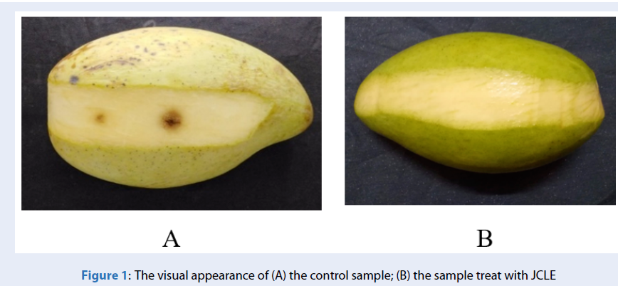
After the 18 day of storage, both treatments had not presented black spots (Table 4). Keeping recording results until the 21 day, the black spots appeared and spread in the following days in the control samples, but the model with JCLE was neither. After 30 days, the blackening started appearing on the JCLE sample's fruit surface. Meanwhile, the blackening of the control samples was inflicted attacking by deep wounds to the flesh fruits (Figure 1). Additionally, the diameter of the control's blackening (6.5 mm ± 0.1) was more than the JCLE sample (2.5 mm ± 0.1). The current study demonstrated that the JCLE could protect mango fruits from the growth of C. gloeosporioides within 27 days.
The experiment also found the physicochemical properties of both treatments were similar after storage at 12 ± 2 for 30 days. Specifically, all mango fruits had a TSS of 10 – 12°Brix and a total acidity of 0.60 – 0.7 %. The mango fruits after 30 days of storage reached a TSS value higher than the fruits before storage, whereas TA was lower (e.g., TSS of 6 – 7°Brix and TA of 0.90 – 1.00%). During the maturation stage, enzymes hydrolyze starch and polysaccharides into simple sugar, while oragnic acids such as citric acid and ascorbic acid may be utilized as substrates for respiratory progression (Palafox-Carlos et al., 2012). On the other hand, the current study obtained the colorimetric values of mango’s pulp: L* from 53.79 to 54.12, a* from - 10.27 to - 9.53 and b* from 37.86 to 38.02; these values were greater than the samples before storage (section 2.1.3). Throughout the ripeness, the increase in the action of the chlorophyllase enzyme caused chlorophyll degradation, and simultaneously, the synthesis of carotenoid led to the reduction of greenness and the gain of yellowness27. In conclusion, the result showed JCLE had little effect on the maturation process of the mango.
|
Treatments |
Time (day) | |||||||||||
|
0 |
3 |
6 |
9 |
12 |
15 |
18 |
21 |
24 |
27 |
30 |
33 | |
|
JCLE |
- |
- |
- |
- |
- |
- |
- |
- |
- |
- |
+ |
+ |
|
Control |
- |
- |
- |
- |
- |
- |
- |
+ |
+ |
+ |
+ |
+ |

The visual appearance of (A) the control sample; (B) the sample treat with JCLE
CONCLUSION
M. calabura leaves extracts had an inhibitory effect on the growth of A. alternata, C. gloeosporioides, F. equiseti, and L. theobromae. The ethanolic solution showed a suitable inhibitory on L. theobromae, A. alternata. The study also found a positive relationship between the antifungal activity and TPC (from 4000 to 40000 mg GAE/kg) of ethanolic extracts from JCLE. At the same applied concentration of 40000 mg GAE/kg, the susceptibility of microbial under the inhibitory effect of JCLE decreased in the following order L. theobromae (75.03%), A. alternata (57.03%), F. equiseti (54.81%), and C. gloeosporioides (53.29%). From the in vivo experiment with mango storage, the ethanolic extract from JCLE could reduce the growth of C. gloeosporioides for the 27 day of storage at a temperature of 12 ± 2C with a concentration of 40000 mg GAE/kg. Therefore, the JCLE has the potential for use as a biological control agent for the control of anthracnose diseases. The results can be used as a new method for preserving and extending the shelf-life of mango fruits.
ACKNOWLEDGEMENTS
The authors wish to thank VLIR-UOS (VN2018TEA478A 103) for financial support for this study.
CONFLICT OF INTEREST
The authors declare no conflicts of interest.
AUTHOR CONTRIBUTION
Nguyen Hoang Phuc Nguyen: carried out the experiment and wrote manuscript. Hoang Quang Binh: developed analytical method and wrote manuscript. Trinh Ngoc Thao Ngan: developed experimental design. Le Trung Thien: Supervisor. Katleen Reas: Supervisor.